|
|
||||
|
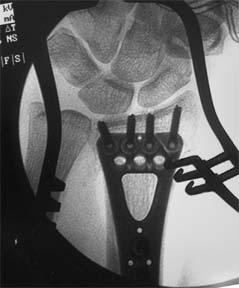
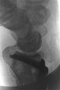
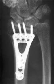
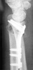
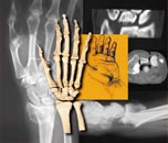
The reduction was considered acceptable. The radius was not out to length, but given the time since the injury (not known precisely, but about a month), it was felt that obtaining full length would not be easily obtainable and not worth an osteotomy. (Note that the facet PA and the facet lateral makes the ulna appear longer than it actually is.) Screw lengths were evaluated and felt to be appropriate, and screws were not in the joint. The patient was seen several times over the next two weeks, and other than calling the office multiple times complaining of severe pain and requesting opioids, she appeared to do well. During the office visits, she requested opioids and complained of many things, including hand numbness (none could be found on exam), inability to sleep, and severe pain (that did not appear supported by her behavior in the office). She returned to work at two weeks on her own initiative. Her pain complaints and requests for Vicodin continued. On followup on November 11, she had full finger range of motion, flexion of 60/82, extension of 42/65, full pronation and full supination, and was continuing to work. She admitted to getting Vicodin from another physician. On January 3, 2006, she called the office complaining of a bump on her hand. We arranged for her to be seen that day. She claimed that the mass occurred immediately after surgery, but prior exams in the office had not demonstrated any masses. Exam showed a soft, fluid-filled mass of 1 x 2 x 3 cm overlying the second and third metacarpals. Although she complained it was painful, it did not appear to be so on exam. She had increased her flexion from 60 to 66 degrees but extension had fallen from 42 to 31 degrees, and she still had full pronation and supination. Her distal radius was not tender to palpation and there was no pain with finger motion, which seemed to rule out a prominent screw. Also, this did not seem to be likely as the mass was over the central aspect of the third metacarpal, not over the distal radius. We suggested observation, but she insisted on an aspiration. This showed typical ganglion type fluid, but the mass recurred while she was still in the offfice. At followup February 16, her flexion was 66/90, her extension had improved to 48/65. She continued to complain of pain shooting up her hand and difficulty grasping things. The mass was unchanged and she was insistent that it be removed. We agreed to remove it.
(6) What is your differential diagnosis?
|
||||
| About Us | Research | Basic Knowledge | What's New | Forum | Guest Professor | Post a Case | eRadius Conference | Patients | Home |