|
|
||||
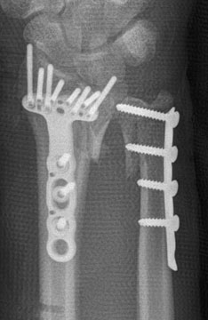
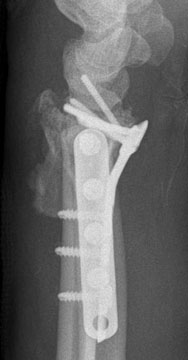
(6) How would you assess the reduction of the radius? The ulna? The xrays obtained by the previous surgeon demonstrate that the reduction of the radius was incomplete on the ulnar side, the "dorsal ulnar corner" fragment was displaced and probably not fixed by any of the screws. This is the challenge that was mentioned on Page 1. There was both proximal displacement and dorsal displacement of this fragment on the initial xrays, but they were hidden once the ex fix was placed. Always look at the initial injury films and, if obtained, traction views, as the size, location, and displacement of the fragments can be seen and their "personality" evaluated. Looking only at the current films may lead you astray. The long screw on the ulnar side of the volar plate indicates to me that the treating surgeon was trying to obtain reduction and fixation on the dorsal ulnar fragment. Locking screws cannot be used in a lag fashion (this is a locking screw) and the fragment still looks displaced. Two challenges that volar plates pose is that you cannot see the intraarticular reduction very well and you cannot easily control the dorsal ulnar fragment. Fluoroscopic views are important and a dorsal incision may be required, and sometimes a dorsal fragment-specific plate as well. Many of the contoured periarticular plates fit a reduced fracture quite well and will keep the screws out of the joint. These plates, with unreduced fractures, however, will not fit and will often place a screw into the joint. This seems to be the case here. The lateral is not a facet lateral and cannot be used to evaluate this question. However, the lateral does show that the radial articular surface is not concentric with the lunate, with an increased AP diameter of the lunate facet, suggesting that the dorsal ulnar corner is not reduced. The proximal location of the lunate on the PA also suggests this. The ulnar fragment was also unreduced and appears displaced proximally. This suggested that the entire DRUJ is proximally displaced, ie, both the dorsal ulnar fragment and the distal ulna. The ulna was fixed with a locking plate, but all the screws were non-locking. In addition, the most distal screw, on these views, was right up to the joint line. Fluoroscopic views or a CT would be able to tell if the screw was in the joint or not. (7) What would be your next step? Further plain films (obliques and facet views) are indicated.
|
||||
| About Us | Research | Basic Knowledge | What's New | Forum | Guest Professor | Post a Case | eRadius Conference | Patients | Home |