|
|
||||
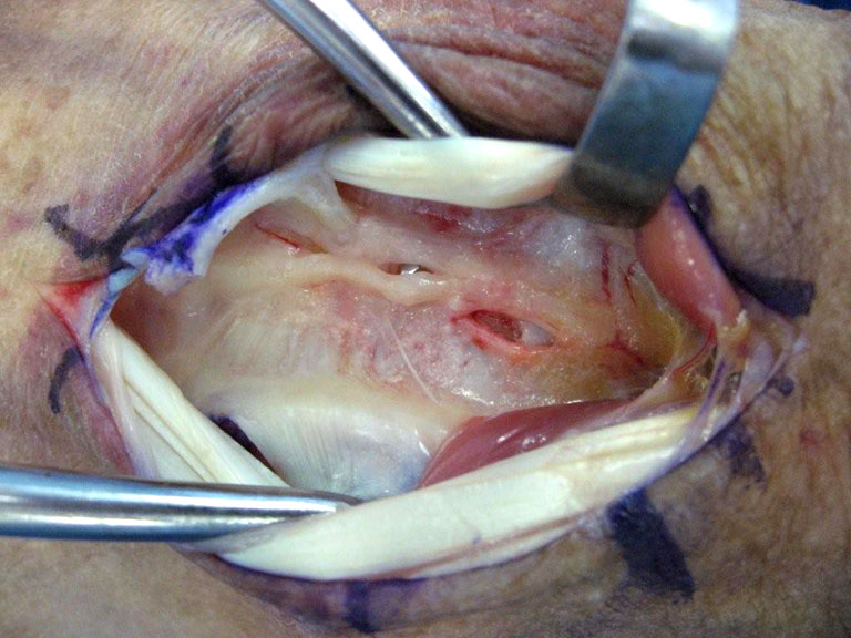
Intraoperative view with the screw tip impinging on the posterior interosseous nerve.
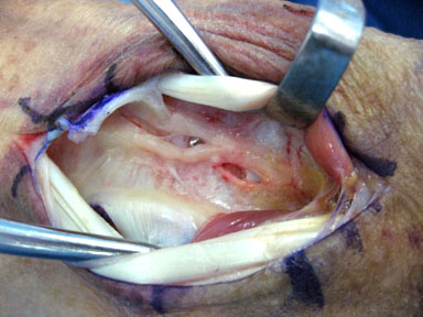
Another intraoperative view with a better view of the screw tip. The screw was removed, with dramatic relief of the patient's pain. She did quite well by 4 1/2 weeks, and did not need further care. Lateral xrays are not a reliable way to evaluate distal screw lengths. Fluorosocopy is better, but screws should still probably stop 2-4 mm below the dorsal cortex to be sure they are not impinging on tendons (see case) or a nerve. There is no need to purchase the dorsal cortex, and no benefit, either. See the paper Complications of Volar Plating for further information. This is the source of the following paragraph: Volar plating requires a new vocabulary and a new awareness of distal radial anatomy. In locked volar plating for DRF’s, in terms of “bicortical purchase”, the “proximal cortex” is the plate and the “distal cortex” is the subchondral bone. There is (1) no need to engage the dorsal cortex, (2) the tendons and the posterior interosseous nerve are in close apposition to the dorsal cortex (<1 mm), (3) it is difficult to determine the precise location of the dorsal cortex and the tendons in relationship to the tip of the screws, and (4) slight pastpointing (~ 1 mm) can lead to tendon injury. It is recommended to have the screw tips 2 to 4 mm short of the dorsal cortex on the lateral xray. For a similar case involving tendon impingement, click here. |
||||
| About UsResearch | Basic Knowledge | What's New | Forum | Guest Professor | Post a CaseeRadius ConferencePatients | Home |