Instructions for the Guest Professor
Note: Guest Professor Cases are by invitation only
To submit you own cases, use Free Cases
1 Pick an interesting and instructive case. It need not be operative or device related. It should illustrate an important concept, technique, or treatment dilemma.
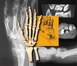
2 Choose the best x-rays: injury film, traction film if indicated, reduction film, special study films if indicated, treatment film, and final film if indicated or available. (Please read the list below carefully. If photomanipulation is new to you, you might want to print this out and check off each step as you go.)
A First, change the image to greyscale. (Even though the image you see on your computer looks like it is in black and white, the camera actually stored it in color, because it is a black and white image. It is still in color. If you do this step first, it will speed up the processing of the later steps.)
B Rotate the image so that the radius is vertical, not slanted, with distal up and proximal down.
C (Optional) Flip the PA image so that the radius is to the left and the ulna to the right and the lateral image so the volar side is to the right. (This helps all images to be standardized.)
D Crop the image to show only the area of interest. In general, only show the wrist from the distal edge of the capitate to about 3 inches down the radial shaft (see prior cases posted on the site).
E Evaluate the quality of the image. Does it need increased contrast? Increased brightness? Modify contrast and brightness to demonstrate the details you are trying to show.
F Convert the digital image to jpg, if not already a jpg, and a resolution of 72 pixels per inch (the resolution of a PC screen). Save it in a Compression Quality Level 5 (medium compression). (Do not do this before cropping, resizing, etc., or you will decrease the quality of the final image.)
G Resize the jpg to a height of 5 inches. This is the same as 288 pixels if you are using 72 pixels per inch. This will make your images fit the screen easily and all the images on the same line will be the same height.
H Remove any patient identifiers from the xray or from the filename. Rename with discriptive names, such as injury_PA.jpg, injury_lat.jpg, post_red_PA.jpg, etc. Do not place spaces in the names, as the Internet cannot accept spaces in filenames the way your computer can; use underscore as shown above.
3 Write short clinical history, approximately 50 words or so. It should be in the format of: "A 26 year old right hand dominant male carpenter fell from a roof approximately 15 feet and landed on his right out stretched arm. He was taken to local emergency room where he was found to be neurovascularly intact. The following x-rays were obtained." The clinical history probably does not need to be anymore extensive than this.
4 Write 3 to 5 provocative, interesting, historical, and/or humorous questions that bring out to teaching point you would like to make.
5 Write a brief summary, a proximally 200 words or less, evaluating treatment of this fracture and the reasons for choosing this treatment. Discuss other treatments that you think might be equally acceptable, and comment on those treatments which you think might be for choices. Cite 1 to 3 references that support your treatment, if possible. (We are trying to build a bibliography section to the Website, and this will be the main way of determining citations.) Propose a few keywords that will help to retrieve this from the Archives (We are trying to build an Archive of prior cases that will be searchable by these keywords.)
6 Be sure to address the questions that you posed.
| 7 Email all of the above to me at: |
 |
|