|
|
||||
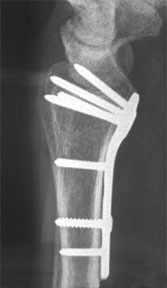
PA and lateral views.
(1) What do you think of the reduction? The reduction looks nearly perfect (the radial length might be just a millimeter short, but really this is quibbling, because the reduction is quite good) and probably is not the cause of her pain. (2) What do you think of the hardware placement (plate placement, screw placement, screw length)? The plate is well placed and is probably not related to the patient's pain. The xrays are not facet views, so screw placement cannot be definitively evaluated. However, the screws are not likely to be in the joint. Screw length can be best evaluated on fluoroscopic views, since the direct lateral can be deceiving. The dorsal cortical surface is not flat in the lateral view, but is a dihedral.
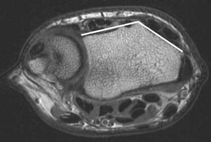
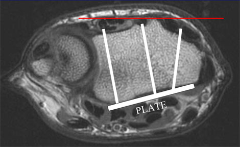
MRI of the distal radius demonstrating the dihedral angle of the dorsal cortex.
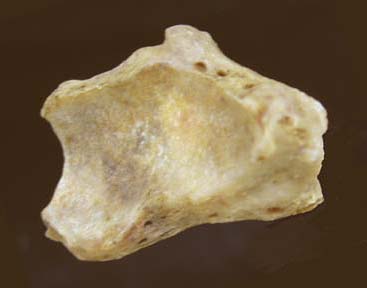
End view of a distal radius demonstrating the dihedral angle of the dorsal cortex. Note also the height of Lister's tubercle.
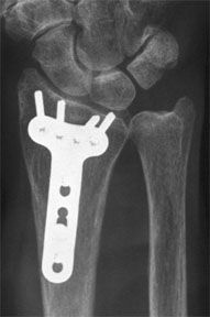
Illustration of a hypothetical volar plate with three screws placed. The red line would be the dorsal surface on the lateral xray. All three screws are too long, yet would appear to be below the dorsal cortex on a lateral xray. The dihedral angle of the dorsal cortex and the height of Lister's tubercle make lateral xrays deceiving with respect to screw length (see Complications of volar plating for a full discussion). The screw lengths, therefore, cannot be evaluated and may be a cause of her pain. (3) How would you work up her pain?
|
||||
| About UsResearch | Basic Knowledge | What's New | Forum | Guest Professor | Post a CaseeRadius ConferencePatients | Home |