|
|
||||
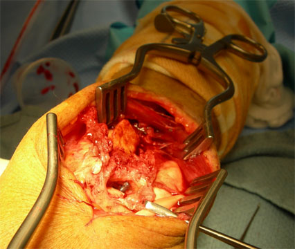
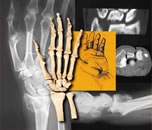
The surgery was, as anticipated, challenging. The first tourniquet time was 2 1/2 hours at 200 mm Hg, down 25 minutes, and a second time of 2 hours at 200 mm Hg was required. The screws were found to be in the radiocarpal joint and in the DRUJ, as shown on the CT.
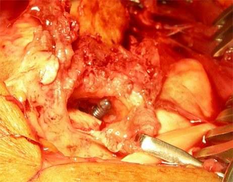
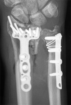
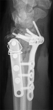
The lunate facet was reduced and the ulnar head fragments were reduced, and each fixed. The distal two ulnar screws were replaced with locking screws, but still were quite unstable. Circlage wires were added. The ulnar fragments were stable to pronation and supination, but not great. (11) How would you interpret these xrays?
|
||||
| About Us | Research | Basic Knowledge | What's New | Forum | Guest Professor | Post a Case | eRadius Conference | Patients | Home |