|
|
||||
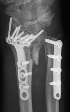
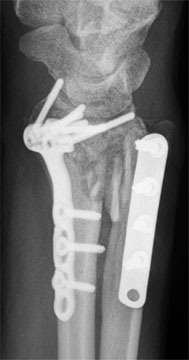
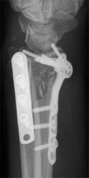
I accompanied the patient to the radiology suite and personally supervised the views. Multiple views were obtained, and additional views were obtained based on an interpretation of the previous views. (8) How do you interpret these views? The ulnar-most radial screw was in the joint, the lunate facet was depressed and angled, the dorsal unlar corner was unstable. The distal-most ulnar screw was suspeciously close to being in the joint, the fragment was displaced proximally and radially. The DRUJ was therefore proximally displaced and not securely fixed. (9) What would be your next step? The patient will need a removal of at least some of the hardware and another attempt at reduction needs to be done. A CT would be needed to better delineate the position of the screws, but in any case, the patient needs surgery. Both should be scheduled in sequence, so as to move forward quickly. The malunion was five weeks out; further delay would make the surgery more difficult. |
||||
| About Us | Research | Basic Knowledge | What's New | Forum | Guest Professor | Post a Case | eRadius Conference | Patients | Home |