|
|
||||
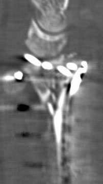
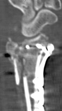
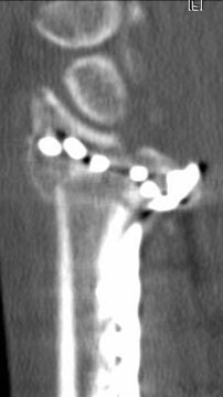
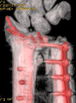
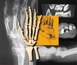
The CT scan showed:
The CT confirmed that the ulnar-most radial screw was in the joint, the lunate facet was angled and displaced, there was an unacceptable diastasis of the articular surface, the distal-most ulnar screw was in the joint, and that the DRUJ was displaced and not securely fixed. As usual, the 3D reconstruction did not add anything. The patient was taken to surgery.
(10) What would be your pre-operative plan? |
||||
| About Us | Research | Basic Knowledge | What's New | Forum | Guest Professor | Post a Case | eRadius Conference | Patients | Home |